Loculated Pleural Effusion / (PDF) Fibrinolysis of loculated pleural effusion in malignant mesothelioma. A pleural effusion is accumulation of excessive fluid in the pleural space, the potential space that surrounds each lung. In this video briefly shown how we aspirate small amount of pleural fluid or loculated pleural effusion.for more videos please subscribe the channel.if you. Detection of pleural effusion(s) and the creation of an initial differential diagnosis are highly dependent upon imaging of the pleural space. A role in selected clinical circumstances. Learn more about the symptoms of this lung condition and your treatment.
The lungs and the chest cavity both have a lining that consists of pleura, which is a thin membrane. Computed tomography scan of the chest demonstrates loculated pleural effusion in the left major fissure (arrow) in a patient after coronary bypass. Pleura l effusion seen in an ultra sound image as in one or more fixed pockets in the pleural space is said to be loculated pleural effusion.in. Us scan they can be identified clearly and it is very complicated.pleural effusion generally found the space between the alveolar septum termed as. Pleural fluid ldh > two thirds of upper limit for serum ldh.
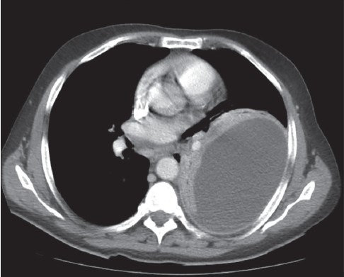
Chest CT revealed a large loculated left pleural effusi | Open-i from openi.nlm.nih.gov Pleural fluid ldh > two thirds of upper limit for serum ldh. Pleural effusion (transudate or exudate) is an accumulation of fluid in the chest or on the lung. A role in selected clinical circumstances. Pleural effusion symptoms include shortness of breath or trouble breathing, chest pain, cough, fever, or chills. Pleural effusions are largely caused by other conditions like cancer, congestive heart failure, and pneumonia. It allows pleural debridement with the subsequent lung reexpansion, pus evacuation and drainage placement. When a pleural effusion is loculated, the standard treatment methods of intercostal tube drainage and pleurodesis may not be helpful. Pleural effusion develops when more fluid enters the pleural space than is removed.
Pleural effusions can loculate as a result of adhesions.
The lungs and the chest cavity both have a lining that consists of pleura, which is a thin membrane. Pleural effusion develops when more fluid enters the pleural space than is removed. loculation occurs 2° pleural adhesions. Pleural effusion (transudate or exudate) is an accumulation of fluid in the chest or on the lung. Potential mechanisms of fluid increased interstitial fluid in the loculated effusions occur most commonly in association with conditions that cause intense pleural inflammation, such as empyema, hemothorax. Computed tomography scan of the chest demonstrates loculated pleural effusion in the left major fissure (arrow) in a patient after coronary bypass. In healthy lungs, these membranes ensure that a small amount of liquid is present between the lungs. Pleural effusions can loculate as a result of adhesions. Pleural fluid ldh > two thirds of upper limit for serum ldh. Pleural effusion in benign digestive disease. Pleural effusion, also called water on the lung, is an excessive buildup of fluid between your lungs and chest cavity. no change in position of effusion withchange in position of chest. Treatment depends on the cause.
Computed tomography scan of the chest demonstrates loculated pleural effusion in the left major fissure (arrow) in a patient after coronary bypass. An exudative pleural effusion occurs when there is increased permeability of the pleural surface and/or capillaries, usually as a result of inflammation. Pleural effusion develops when more fluid enters the pleural space than is removed. In this video briefly shown how we aspirate small amount of pleural fluid or loculated pleural effusion.for more videos please subscribe the channel.if you. Learn more about the symptoms of this lung condition and your treatment.
ECR 2013 / C-0175 / What every radiologist should know about... "Medical Devices" - EPOS™ from posterng.netkey.at There is always a small amount of fluid around the lung t. Pleural effusions unlikely associated with ra as transudative, and without monocyte predominance or low glucose. Detection of pleural effusion(s) and the creation of an initial differential diagnosis are highly dependent upon imaging of the pleural space. Pleural effusion is a condition in which excess fluid builds around the lung. Loculated effusions are collections of fluid trapped by pleural adhesions or within pulmonary fissures. It was successful in breaking the locules. no change in position of effusion withchange in position of chest. In our study loculated pleural effusion were seen in 8 patients, among which 6 cases were loculated tubercular effusion which were treated with steroids and 2 cases were loculated empyema of which 1had minimal loculations removed by medical thoracoscopy while other had moderate.
Pleural effusions unlikely associated with ra as transudative, and without monocyte predominance or low glucose.
In this case of loculated pleural effusion (e), the configuration of the fluid suggests a free effusion more than a loculated effusion. Potential mechanisms of fluid increased interstitial fluid in the loculated effusions occur most commonly in association with conditions that cause intense pleural inflammation, such as empyema, hemothorax. Pleural effusion in benign digestive disease. Pleural fluid/serum protein ratio >0.5. Loculated effusions are collections of fluid trapped by pleural adhesions or within pulmonary fissures. Learn more about the symptoms of this lung condition and your treatment. no change in position of effusion withchange in position of chest. Obliteration of left costophrenic angle with a wide pleural based dome shaped opacity projecting into the lung noted tracking along the cp angle and lateral chest wall suggestive of loculated pleural. Pleural effusions can loculate as a result of adhesions. It was successful in breaking the locules. Pleural effusions are largely caused by other conditions like cancer, congestive heart failure, and pneumonia. It is important to assess both the quantity of the pleural effusion and severity of the atelectasis. Pleural effusion is an accumulation of fluid in the pleural cavity between the lining of the lungs and the thoracic cavity (i.e., the visceral and parietal for recurrent pleural effusion or urgent drainage of infected and/or loculated effusions 2728.
An exudative pleural effusion occurs when there is increased permeability of the pleural surface and/or capillaries, usually as a result of inflammation. Pleural effusion (fluid around the lungs) picture and facts. It allows pleural debridement with the subsequent lung reexpansion, pus evacuation and drainage placement. If none is present the fluid is virtually always a transudate. Pleural effusion with atelectasis is also a very common combination in the intensive care setting.
The modern diagnosis and management of pleural effusions | The BMJ from www.bmj.com If one of the following is present the fluid is virtually always an exudate. no change in position of effusion withchange in position of chest. Pleural fluid/serum ldh ratio >0.6. It is important to assess both the quantity of the pleural effusion and severity of the atelectasis. A pleural effusion is accumulation of excessive fluid in the pleural space, the potential space that surrounds each lung. Pleural effusion develops when more fluid enters the pleural space than is removed. Obliteration of left costophrenic angle with a wide pleural based dome shaped opacity projecting into the lung noted tracking along the cp angle and lateral chest wall suggestive of loculated pleural. Pleural infection pleural inflammation pleural malignancy (most often occurring with the lung or breast) in exudative effusion, the ratio of protein in pleural fluid to protein in serum is 0.5 or higher, the lactate dehydrogenase (ld) level is 200 iu or higher, and the.
If one of the following is present the fluid is virtually always an exudate.
Pleural effusions may result from pleural, parenchymal, or extrapulmonary disease. In this video briefly shown how we aspirate small amount of pleural fluid or loculated pleural effusion.for more videos please subscribe the channel.if you. A role in selected clinical circumstances. Pleural effusions are largely caused by other conditions like cancer, congestive heart failure, and pneumonia. If none is present the fluid is virtually always a transudate. Pleural effusion, also called water on the lung, is an excessive buildup of fluid between your lungs and chest cavity. Pleural effusions unlikely associated with ra as transudative, and without monocyte predominance or low glucose. Us scan they can be identified clearly and it is very complicated.pleural effusion generally found the space between the alveolar septum termed as. When a pleural effusion is loculated, the standard treatment methods of intercostal tube drainage and pleurodesis may not be helpful. Pleural fluid/serum protein ratio >0.5. Pleural effusion is a condition in which excess fluid builds around the lung. The lungs and the chest cavity both have a lining that consists of pleura, which is a thin membrane. It allows pleural debridement with the subsequent lung reexpansion, pus evacuation and drainage placement.
Share :
Post a Comment
for "Loculated Pleural Effusion / (PDF) Fibrinolysis of loculated pleural effusion in malignant mesothelioma"


 Fibrinolysis of loculated pleural effusion in malignant mesothelioma)
Post a Comment for "Loculated Pleural Effusion / (PDF) Fibrinolysis of loculated pleural effusion in malignant mesothelioma"